Lipid Simulations


Biological Membranes


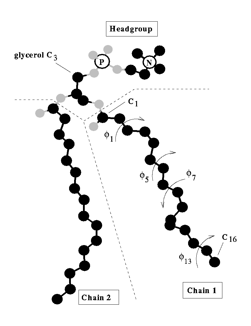
Lipids are the building blocks for biological membranes. They are characterized based on three basic components: (1) a dipole headgroup that is hydrophilic or "water loving", (2,3) and two hydrocarbon chains that are hydrophobic or "water hating". These chemical properties give the lipid bilayer a preferred construct: the headgroups stay near the water and the chains hide in the interior of the membrane. Most lipids investigated by the lab are phospholipids characterized by the presence of phosphate (P) and choline (N) subgroups in the headgroup. The tails are classified by the number of carbons, and position and number of carbon-carbon double bonds which increase flexibility. A glycerol backbone links the headgroup and hydrocarbon tails.

Courtesy of Richard M. Venable, CBER, here is a sketch of a DPPC molecule with several dihedrals labeled on the sn-1 chain. (click for video)
SDPC bilayer. Feller et al, Biophys J. (1997).


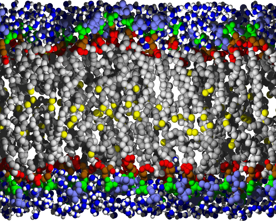
Biological membranes are composed of two layers of lipids organized into sheets. They form a nearly impermeable barrier to water-soluble molecules, especially ions. Membrane-bound proteins interact with lipids to establish a controlled mechanism for signaling across the barrier and for diffusion of material through the barrier. Bilayers can also act as an anchoring point for proteins.


Membrane and Lipid Simulations


Click to download .pdb files where available.


POPC bilayer supported on nanoporous silicon slab.  Roark, & Feller, Langmuir (2008).
POPC bilayer supported on nanoporous silicon slab. Roark, & Feller, Langmuir (2008).
DMPC bilayer with 4 N-Ras peptides.  Vogel et al, BBA-Biomembranes (2010).
DMPC bilayer with 4 N-Ras peptides (orange backbone, yellow lipid modifications). Vogel et al, BBA-Biomembranes (2010).
All-trans retinylidene with double bonds labeled orange.  Feller et al, Biophys J. (2011).
All-trans retinylidene with double bonds labeled orange. Mertz, Lu, Brown, & Feller, Biophys J. (2011).
DHA conformations sampled from lipid bilayer simulation, Feller et al, JACS (2002). DHA conformations sampled from lipid bilayer simulation, Feller et al, JACS (2002). DHA conformations sampled from lipid bilayer simulation, Feller et al, JACS (2002).
DHA conformations sampled from lipid bilayer simulation, Feller et al, JACS (2002).
Rhodopsin embedded in a stearoyl docosohexaenoyl PC bilayer. Feller et al, JACS (2003).
POPC bilayer.  Armen, Uitto, & Feller, Biophys J. (1998).
POPC bilayer. Armen, Uitto, & Feller, Biophys J. (1998).
DPPC bilayer in the fluid phase. Feller, Venable, & Pastor, Langmuir (1997).
Prefered interfacial location of ethanol within a POPC lipid bilayer, Feller et al, Biophys. J (2002).


Back to home page.